Connect with Dr. James Fondriest DDS, FICD, Dentist in Lake Forest, IL

Dr. James Fondriest DDS, FICD, - Dentist

5.0 based on 5 reviews

1 Rating out of 5 stars

847-234-0517 View Phone

560 Oakwood Avenue, #200

Lake Forest, IL 60045
Google Map

Like Dislike
Dr. James Fondriest is ranked #1 out of 3 Lake Forest Dentists Rating 5.0 stars based on 5 reviews $$
Health Spa Facilities
First Class Service
Painless Procedures
Superb Results
Moderate Cost
Accepts New Patients:
Meet the Doctor

Dr. Fondriest is a curriculum author, and a senior lecturer for the Post Graduate Cosmetics Curriculum at the respected Pankey Institute* in Key Biscayne, Florida. His Lake Forest office focuses on cosmetics, reconstructive, and implant dentistry. He lectures internationally and has published many articles on cosmetic makeovers, creating natural looking dentistry, and implants. Some of his professional memberships include the American Dental Association, Academy of Fixed Prosthodontics, and the American Academy of Cosmetic Dentistry. His office is open to visiting dentists from around the world. Jim is a past President of the North Suburban Dental Society of Chicago. He has served on the State Dental Board in Illinois and has rece

Dental Practice Overview based on patient reviews
    Office Facilities & Resources

    Before & After Cases On Website


    Before & After Cases On Website


    Link
    Office Photos
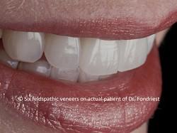
    Feldspathic veneers by Dr. Fondriest
    Feldspathic veneers by Dr. Fondriest
    Special Procedures

    Leading Chicago Northshore Cosmetic And Implant Center

    DR. FONDRIEST maintains Lake Forest Dental Arts, a full-time private practice with a primary focus on comprehensive esthetic restorative dentistry. He is also a nationally recognized speaker & educator on the topics of cosmetic dentistry and prosthodontics and the latest advances in these rapidly evolving disciplines. A well published author, Dr. Fondriest has a strong commitment to providing the highest quality of dental care, along with developing sound educational programs for general dentists aspiring to do more complicated dental implant procedures, smile makeovers, porcelain veneers and crowns. Having began his career as a general dentist; Dr. Fondriest accepts and maintains a patient base for which he provides routine general dental care. "Many of my patients are people that I grew up with in Lake Forest, as well as their families. I look forward to the opportunity to share in the lives of my patients, many of them for decades, says Dr. Fondriest. I am proud to say that I still have many of the patients that were in the practice when I took it over. Dr. Fondriest lives and practices in Lake Forest, Illinois. He and his wife Diane have two children and a hound dog. Dr. Fondriest has been very active in his community having served on the boards of United Way, Lake Forest Rotary, Gorton Community Center and his church. He is an avid amateur photographer and enjoys jogging, eating well, and adventure trips. Dr. Fondriest is an active member of numerous professional organizations, including the American Dental Association, the American Academy of Cosmetic Dentistry, the American Academy of Fixed Prosthodontics, the Academy of Osseointegration implant society, and the American Equilibration Society. Dr. Fondriest is the past President of the North Suburban Dental Society of Chicago. He was elected to the Board of Directors of the Illinois State Dental Society and has been awarded honorary fellowships in the prestigious International College of Dentis


    Link
    Articles

    Choosing Dental Implants Dec. 13, 2011, 2:11 a.m.

    Lake Forest Dental Arts Leads the way in Dental Implants

    Modern dental implant treatment has been used for more than 25 years. In the last 10 years, it has become increasingly popular and more affordable. Dental implants are a high quality, durable, and esthetically pleasing treatment to replace missing teeth or to secure a denture, partial, or bridge.

    Many people suffer from tooth loss because of extensive dental decay, periodontitis (advanced gum disease), or trauma. An estimated 25% of Americans over age 65 have lost all of their teeth, according to the CDC. Tooth loss contributes to poor diet, low self-esteem, and an inability to speak clearly. Fortunately, people do not have to live without teeth. Whether you’re missing one tooth or all of your teeth, Dr. Fondriest offers practical, long-term solutions sure to make you smile.

    Dental implants provide the patient with a fully functioning and natural looking dental prosthetic, without relying on the gums or surrounding teeth for support. Partials and bridges do rely on adjacent teeth. Bridges feature crowns, secured to healthy teeth, to hold the prosthetic in place. Partials have clasps that attach to neighboring teeth. Placing more weight and work on healthy teeth can lead to more dental damage in the long run. Dentures are made to rest on the contours, or ridges, of the gums. Over time, the natural ridges wear down and dentures become more prone to slippage, looseness, and poor fit.

    At Lake Forest Dental Arts, we want to preserve your smile and restore your confidence, while using the marvels of modern dental technology and science. Dr. Fondriest provides many options for dental implants, and based on your case, he’ll recommend the optimal choice. With proper care, your dental implants can last a lifetime.

    To find out if how dental implants can change your life, schedule your consultation with Dr. James Fondriest today. Call 847-234-0517. Lake Forest Dental Arts serves patients of North Chicago and surrounding areas.


    Link

    MINIMAL PREPARATION VENEER CASE SELECTION PROCESS Dec. 4, 2010, 6:34 a.m.

    MINIMAL PREPARATION VENEER CASE SELECTION PROCESS

    Fondriest J, Roberts M. Prepless Veneer Case Selection. Inside Dentistry. March 2010; 6: 3643.

    by James Fondriest, DDS; Matt Roberts, CDT

    No- or minimal-preparation veneers offer both benefits and limitations.

    No synthetic restorative material used to reproduce natural tooth structure can match the combination of ideal qualities of functional strength and optical or esthetic display that exists in nature. Maintaining as much natural tooth structure as possible is a goal when doing restorative dentistry, especially when done for elective purposes. While less tooth reduction is a desirable goal, there are times when more reduction better serves the overall restorative agenda.

    Evaluation Process for Minimum Preparation Veneer Candidates

    It is critical to carefully appraise the patient’s pre-existing condition, tooth position, and dentition color as well as functional envelope, phonetic components, and the patient’s perceived goals of treatment before deciding the possible modalities of treatment. A comprehensive examination with a complete set of records and photographs should be taken to evaluate the interaction of function and determine the esthetic result desired. Mounted models can be compared with the facial photographs to analyze the desired changes to be made.

    Additive vs Subtractive Dentistry

    The functional and esthetic components of restoring teeth include planning the ideal alignment, shape and contour, surface morphology, incisal edge positions, and the opposing functional surfaces. The existing teeth can either be moved orthodontically into position or the tooth contours are modified by subtraction or addition. There has been a trend in restorative dentistry toward less invasiveness by means of tooth reduction and more common partnering with orthodontists with the goal of less tooth reduction or subtractive dentistry. Orthodontic and periodontal solutions to biologic and esthetic problems should be accomplished before planning definitive restorative solutions.

    Diagnostic Wax-Up

    The patient’s models and photographs are used to evaluate any supplemental restorative repositioning of teeth beyond orthodontics necessary to achieve the treatment goals (Figure 1 and Figure 2. The photographs are faithfully oriented relative to the facial midline to properly assess the occlusal plane and tooth axial alignment issues. Line drawings of the proposed restorative treatment are created on the photographs (Figure 3) which assist the technician in choosing landmarks for a wax rendering of the treatment agenda. An "additive-reductive wax-

    Figure 1. This patient’s goals were to straighten his smile, close diastemas, and to make his teeth show more in his smile. There was interest in brightening the teeth if it could be done with the restorations. This patient would not consider orthodontics as an option.

    Figure 2. Patient had little time to invest in treatment and little desire to extend treatment beyond a short time commitment (his wedding date). He wanted to know how little drilling could be done with the greatest impact on his smile. He was highly aware, well versed on treatment options available in the current market, and wanted involvement in treatment decisions.

    up" is done (Figure 4) where contour is added in wax or removed from the stone models of the patient’s existing dentition using the facial photographs to orient the procedure. This process produces a template of the ideal esthetic form for the patient, and from this an intraoral silicone matrix can be fabricated for use in provisional fabrication and intraoral mock-up evaluation. If reductive changes have to be made to reach the ideal tooth position, those areas at least will require some tooth preparation, without which the patient is forced to accept a compromised treatment outcome.

    Mock-Up Evaluation of Additive-Only Esthetic Outcome

    For patients who visually may be candidates for no-preparation veneers, an additive-only wax-up can be done; after a silicone matrix of this wax-up is created, the shape and position of this wax-up can be tested in the patient’s mouth by filling the matrix with a material such as Luxatemp® and placing it over the existing dentition to create an intraoral mock-up (Figure 5). An intraoral mock-up may also be accomplished with direct bonding. If an esthetically pleasing outcome can be accomplished by additive procedures only, the case is one step closer to qualifying for very conservative veneers with little or no preparation.

    Functional and Phonetic Evaluation of Mock-Up

    If the mock-up results are esthetically pleasing, then a phonetic evaluation should be undertaken. After an evaluation of "f," "v," and "s" sounds is completed, the overall form of the teeth should be looked at. Excessive incisal thickness of maxillary anterior teeth is quite common after an additive mock-up, which may lead to the necessity of lingual preparation in some cases. Most esthetic changes in the anterior incisors will have functional ramifications as well. Canine guidance, crossover, and protrusive positions must be evaluated while the patient is wearing the mock-up. Changes in the incisal-edge positions of the maxillary incisors almost always create the need for modifications to the mandibular incisors to maintain a comfortable and atraumatic guidance.

    Changing Brightness with Low-Preparation Veneers

    Low-preparation veneers are not a good choice when attempting to brighten teeth. By virtue of the design of these veneers, they are thin; usually less than 0.5 mm thick. This minimal thickness will have minimal effect on the brightness unless they are made with opaque porcelain. Using opaque porcelain has its own drawbacks, mainly the lack of natural translucency and an unnatural appearance. The color of the existing dentition must be considered and balanced with the final tooth shade desired by the patient. The more change in color desired, the thicker the ceramic layer will need to be to provide adequate filtering of underlying

    color. If the intraoral mock-up using a translucent material has the needed thickness to achieve the desired

    Figure 3. Significant clinical findings included a bruxing habit with moderate to heavy wear for age, gingival recession, occlusal plane cant, lack of symmetry between maxillary incisal edges and lower lip, diastemas, alignment issues, otherwise healthy and unrestored teeth.

    Figure 4. Diagnostic wax-up was employed to assess the amount of subtractive-additive changes needed to achieve treatment goals.

    Figure 5. Luxatemp mockup was done so the patient could see and feel the proposed new tooth orientations. The mock-up procedure often offers a very positive response from the patient increasing engagement and treatment compliance.

    color change, the case can proceed with no preparation or minimal preparation. If, when evaluating the mock-up, there is chromatic show-through of the underlying dentition and it is producing an undesirable color gradient, the underlying tooth structure will need to be reduced to a level that provides an adequate amount of room for the ceramist to filter out the undesired effect with the porcelain while maintaining natural vitality and translucency. The depth of preparation that is necessary completely depends on the severity of the discoloration and the amount of color change being attempted in the treatment.

    Path of Insertion

    If after evaluating for position, form, function, phonetics, and color, the patient is still a candidate for no- or minimal-preparation veneers, the path of insertion and potential undercuts that may limit access to the areas of the teeth requiring augmentation now needs to be determined. The stone models and the additive diagnostic wax-up should be used to determine which areas of the teeth are going to be involved in the restorations. All areas covered in wax will need to be covered with the restorations, and will need a common path of insertion. A study model of the patient’s pre-existing dentition can be placed on a surveyor in the laboratory and each restoration can be checked for path of insertion to all surfaces restoratively involved, and undercuts can be identified and marked. If a surveyor seems excessive, a simple visual check to identify undercuts on the stone model can work well for skilled operators. All areas that are identified as being undercut will need to be addressed with additive bonding or tooth reduction. If at this time there are no apparent undercuts, or they can be solved with additive bonding to allow a path of insertion, the patient is a candidate for "no-prep veneers."

    Minimal Preparation Philosophy

    There are many attractive reasons why the practitioner would want a case to be minimal preparation. When minimal tooth reduction is done, there is less preparation time and, thus, a shorter appointment. Anesthetic is either not needed or it is used sparingly. The fact that no or limited tooth structure is removed means intermediate provisional restorations are not required. Although accurate impressions are just as critical with minimal-preparation techniques, the invasiveness and difficulty of the impression technique is diminished. With less preparation, there is a likelihood of retaining more enamel, which increases bond strength and the long-term integrity of the margins. This adds to the durability and longevity of the final restorations. If, after completion of the evaluation steps above, there are a few areas of limitation that are not global in nature, this is where minimal preparation comes into play.

    Illusions of Reversibility

    The patient was highly motivated to have as little tooth reduction done as possible while achieving as many of his treatment goals as possible. Not knowing initially if he would like the added length and changed shapes of his planned restorations, the patient gained comfort from the possibility of full reversibility if the underlying teeth were untouched. The patient acknowledged that the treatment was being done mainly for vanity or cosmetic reasons. If, after completion, it was determined this was all a mistake or the result failed to live up to expectations, then it would be a highly desirable option to go back to the way he was initially. While true in theory, it is not exactly true in practice, as it is very difficult to remove bonded veneers cleanly. The layers of porcelain, bonding material, and tooth structure are difficult to distinguish when using rotary instruments at high speed and with a water spray. Unintentionally, some tooth structure would be lost, and surface texture and morphology would be forever changed. Although this is definitely not a reversible procedure, the likelihood of losing only a minimal

    Figure 6. Image taken after some minimal preparation. Some sharp corners were subtly rounded, line of draw undercuts reduced, and small chamfer margins were prepared.

    amount of tooth structure was still very attractive to the patient (Figure 6).

    Preparation Benefits

    Small areas of tooth preparation are accomplished to eliminate the problems with path of insertion, color, and function while maintaining an overall philosophy that is very conservative of the existing natural dentition. This allows the completion of treatment that delivers an uncompromised esthetic and functional result for the patient and maximizes the amount of retained natural dentition. If the limitations seem to be of a more global extent, then traditional veneer or crown preparation may be necessary to treat that particular patient.

    There are significant benefits to be gained with tooth preparation. Because of the lower flexural strength of porcelain, wrapping porcelain around sharp corners creates a greater likelihood of crack propagation. Simply rounding out the sharp corners and edges of the underlying teeth will improve the durability of the restorations.

    Rough surfaces on teeth tend to attract stains and debris. The margins of the newer porcelains used for veneers have minimal thicknesses that are measured in tenths of millimeters, but adaptation to the underlying tooth structure (the fit) must still be accurate to minimize the resin component at the margin interface. Small undercuts in marginal areas which result in overhanging ceramic that is not welladapted to existing tooth structure can create significant resin thickness at the margin. In microscopic and microbiotic terms, this can create huge areas that are difficult to clean. The thicker the margin is, the more it will stain. These visible and undesirable stains can appear within 6 weeks or less of delivery. This will happen sooner if the margin is not wellsealed with luting material. A large percentage of patients have gingival recession over time, leaving the staining margins even more visible and unattractive. Just a shallow chamfer preparation at the planned margins of the restoration allows a smooth marginal transition, healthy tissue, and decreased likelihood of marginal staining Figures 79).

    Laboratory Perspectives

    The restorations used for this type of treatment require special skills and the use of specific material in the laboratory. From a laboratory perspective, ease of fabrication is directly proportional to the amount of tooth reduction achieved by the dentist; the more reduction, the more material options available and the easier the fabrication of the restorations. The most difficult cases are the "no- prep" cases; although a minimal-preparation treatment approach can be easier than a no-preparation approach, the minimal thickness of material still makes these cases much more difficult to fabricate than a traditionally prepared veneer case that is prepared to transition smoothly from a minimum thickness of 0.5 mm to maximum thicknesses in the range of 1.5 mm.1 Minimal-preparation veneer porcelain can have thicknesses as little as 0.1 mm in some areas and over 1 mm thick on the same restoration with more abrupt transitions from thin to thick areas, requiring careful selection of ceramic material to deal with the underlying color and translucency levels of the preparations.2

    Figure 7. One week post-delivery. Shallow chamfer at margins allowed for healthy tissue but no cervical reduction was done. A common characteristic of minimum prep cases is overcontoured "bulbous" cervical especially with the canines.

    Figure 8. One week post delivery of minimal preparation veneers

    Figure 9. close-up view of pressed minimal preparation veneers showing that natural surface morphology can still be achieved with porcelain that is less than 0.5mm thick

    Materials

    There are two traditional porcelain options for making veneers. A long-standing technique that can yield beautiful results is to stack feldspathic porcelain on either a platinum foil or refractory die. Advantages include the ability to vary the opacity and chroma levels in different parts of each individual restoration as needed. The main limitation to these restorations is the very fragile nature of thin feldspathic veneers, which can crack easily during fabrication and placement and exhibit a flexural strength in the range of 85 MPa.3 Today, it is possible to fabricate very thin pressed veneers using high-translucency lithium-disilicate ceramic material, which has a flexural strength of 400 MPa, thus eliminating the delicate handling necessary with feldspathic material.4 This material can be pressed more thinly than usual feldspathic veneers can be fabricated (to 0.1 mm), which works well over desirably colored tooth structure.

    Lab Fabrication

    Lithium disilicate was selected for this case because of its ability to be pressed into very thin restorations; in this case, 0.2 mm in many areas of the facial portions of the veneers. The 400 MPa flexural strength of this material also decreases the risk of fracture during insertion with the complex path of insertion necessary with these very thin veneers.5 High-translucency, enamel-like ingots were used in the lost-wax pressing process, which eliminated the need for layering in the gingival third. The incisal third was cut back slightly from the facial to allow layering of incisal effects (Figures 10-12)

    Arguments for More Preparation

    A presumption made earlier in this article was that it is easier for the dentist to do minimal-preparation veneers. There is a good argument for the preparation of low-preparation veneers being more technique-sensitive than normal-preparation veneers.6 This is especially true when the dental alignment is not perfect, diastemas exist, or there is a lot of gingival recession with large interproximal embrasures (interproximal space devoid of tissue).7 With well-aligned teeth that have tissue filling the interproximals, there is less wrap required to hide the margins. The porcelain can be more uniform in thickness and the anatomy of the teeth is only changed a little (buccal and incisal embrasures tend to be smaller). When teeth are misaligned or when more interproximal cavosurface is exposed, the porcelain needs to wrap more interproximally to fill the space so that all interproximal margins are tucked out of view. Many of these surfaces bend around more than 180° of the circumference, thus yielding undercuts and line-of-draw issues during fabrication and placement of the restorations. Laboratory fabrication issues escalate substantially. Trying to treat cases that exhibit path of insertion problems related to diastemas or rotations without adequate preparation can leave the ceramist in the undesirable situation where undercuts prevent adequate interproximal path of insertion and leave the ceramist with a choice of having to move the proximal contacts too far to the facial, resulting in a bulky appearance,or blocking out the undercut and extending the margins interproximally to allow ideal contour, but resulting in an open margin where the undercut was blocked out.

    Advantages of Normal Preparation Over Low Preparation

    1. Subtractive capabilities allow for more natural tooth contours
    2. More control of buccal/lingual placement of incisal edges

    LABORATORY WORK (10. & 11.) Minimal facial/incisal layering produced desired translucency while leaving the maximum amount of pressed lithium disilicate intact(12.) Finished veneers on solid model.

    1. Better control of midline alignment
    2. Shade change without creating unnatural opacity
    3. Less bulky emergence contours, especially on the cuspids
    4. Easier placement with multiple line of draws
    5. Less technique-sensitive for the dentist and technician
    6. Easier to hide margins
    7. Decreased tendency toward more closed/smaller buccal and incisal embrasures
    8. More symmetrical shapes, especially of lateral incisors
    9. Nicer surface textures
    10. Nicer incisal characterization

    The Ideal Patient

    The ideal candidate for very conservative treatment is one with a slightly underfilled buccal corridor; slightly lingualized, small maxillary anterior teeth; closed or almost closed contacts; relatively even spacing of teeth; no gingival recession with gingival tissue filling interproximal areas; and no severe discoloration. Patients with full, slightly overdominant teeth are not good candidates, neither are severely tetracycline-stained cases.8 Patients exhibiting loss of interproximal soft tissue, crowding, and rotations require careful screening described earlier and usually require orthodontics before veneering.9 Patients exhibiting excessive spacing require orthodontic treatment before veneering as well.10 The patient population that has all of these characteristics is small.8

    Conclusion

    The amount of tooth reduction should be matched to the goals and priorities of the patient. There are positives and negatives with both heavy and light tooth preparation that significantly affect the quality of outcome. Low-preparation veneers are a good choice when modest improvement in symmetry and tooth alignment is desired as well as making a more uniform surface morphology. The best cases for the minimal-preparation design are those with properly aligned teeth without diastemas, gingival recession, or papillae loss. The minimal- preparation veneer is not a good choice when the goal is to greatly change value or brightness.

    If the case selection is not ideal, minimal-preparation veneers can be more technically challenging for the practitioner and the technician than conventional veneer designs. The practitioner is responsible for controlling undercuts, creating a line of draw, and hiding margins. Significant improvements in outcome can be achieved with small amounts of tooth preparation to create a margin, open buccal and incisal embrasures, ensure a line of draw, and round out sharp edges.

    The philosophy of doing conservative dentistry is a noble goal but it should noted that conservative does not mean limited preparation but, rather, preparing the least amount of tooth structure needed to achieve the goals of the case.

    References

    1. Puri S. Techniques used to fabricate all-ceramic restorations in the dental practice.
    Compend Contin Educ Dent. 2005;26(7):519-525.

    2. Calamia JR. Etched porcelain facial veneers: a new treatment modality based on scientific and clinical evidence. N YJ Dent. 1983;53(6):255-259. 3. Friedman MJ. A 15-year review of porcelain veneer failure—a clinician’s observations.

    Compend Contin Educ Dent. 1998;19(6):625-632.

    4. Ritter RG, Rego NA. Material considerations for using lithium disilicate as a thin veneer option. Journal of Cosmetic Dentistry. 2009;25(3):111-117. 5. DiMatteo A. Prep vs. no-prep: the evolution of veneers.

    Inside Dentistry. 2009;5 (6):72-79.

    6. Ferrari M, Patroni S, Balleri P. Measurement of enamel thickness in relation to reduction for etched laminate veneers. Int J Periodontics Restorative Dent. 1992;12(5):407-41 3. 7. Calamia JR, Calamia CS. Porcelain laminate veneers: Reasons for 25 years of success.

    Dent Clin NAm. 2007;51:399-417.

    8. Strasseler HE. Minimally invasive porcelain veneers: indications for a conservative esthetic dentistry treatment modality. Gen Dent. 2007;55(7):686- 694. 9. Javaheri D. Considerations for planning esthetic treatment with veneers involving no or minimal preparation.

    JAm Dent Assoc. 2007;138(3):331-337.

    10. Jacobson N, Frank CA. The myth of instant orthodontics: an ethical quandary. J Am Dent Assoc. 2008; 1 39(4):424-434.

    About the Authors:

    James Fondriest, DDS Private Practice

    Lake Forest, Illinois

    www.lakeforestdentalarts.com

    Matt Roberts, CDT CMR Dental Lab

    Idaho Falls, Idaho

    www.teamaesthetic.com


    Link

    Masters Technique Level: Shade Matching the Single Central Dec. 4, 2010, 6:29 a.m.

    James F Fondriest, DDS560 Oakwood Avenue, Suite 200Lake Forest, IL 60045 
    Abstract 
     
    There are very few things our client patients can evaluate our talents on clinically.  They do not really know if our margins fit or the occlusion we have designed for them is adequate or appropriate.  They do know however if our restorations match.  Therefore it is important for the dentist/technician team to develop their skills to allow for the faithful reproduction of nature in our restorations.  Developing our skills so that we can communicate exactly what is seen in the mouth will allow the laboratory technician to produce a more lifelike replica.  This paper suggests a protocol for the evaluation of the most important parameters of a match and for the photographic communication of these parameters between the laboratory and the dentist practitioner.  
      Introduction              Accurately reproducing the unique characteristics of a single maxillary central so that the artificial replacement is perceived as “natural” can be the biggest challenge in restorative dentistry.  This challenge comes at several levels.  First, the practitioner needs to have an understanding of what factors go into achieving a good match and some basic knowledge of the nomenclature of light science in order to communicate what is seen.  Secondly, the practitioner is responsible for creating a protocol to accurately assess what is happening when light hits the surface of the tooth to create its visual appearance.  There are ways to increase what we see visually or photographically in the mouth lessening metamerism, afterimages, and other visual distortions.  Thirdly, the practitioner needs to develop written, graphic, and photographic communication devices that are more comprehensive and less confusing.  For example, we have no common dental standard for communicating the degree of translucency, hypocalcification of enamel, or varying degrees of surface luster.  If we had to describe in great detail what we see in the teeth in Figure 1 without photography, how many words would it take to deliver a non-confusing synopsis?  And lastly, the laboratory needs to develop itself along with the practitioner partner because all of the levels of communication conveyed to the lab must also be recognized and understood in the photographs.

    Figure 1

    How easy would it be to describe these teeth in written form?

    It is important to realize that matching the hue and chroma is fifth or sixth in importance on the list of things to match when constructing a prosthetic replacement 1.  You have to be fairly close to someone to detect subtle differences in hue; yet surface morphology, value, and opacity disparities can be seen from four or five feet away or more.  Disparate tooth silhouettes or perimeter shapes of the teeth can be seen from even 10 feet away. The order of importance while matching a single maxillary central is: 1.    silhouette or perimeter shape2.   

    Figure 2

    A high percentage of light that hits at 900 will return to the viewer while light that hits a surface at an oblique angle will be deflected away from the viewer.  An observer only sees an object when light comes from that object.  Surfaces that are perpendicular to us send the most light back to us.  

    surface morphology and texture3.    value4.    translucency or opacity5.    chroma6.    hue Shape, Morphology, and Texture The appearance of teeth is mostly determined by how light interacts with its curved and varied surface.  The perimeter shape and the morphology of the buccal surface have the greatest affect on the appearance of teeth because they determine how the majority of light is reflected. An observer only sees an object when light comes from that object.  Surfaces that are smooth and perpendicular to us send more light back to us (see Figure 2).  The reflective surfaces of the tooth will not return significant light to our eyes if they are not perpendicular to our eyes even if they are highly polished.  Figure 3 shows an example of in this case sand which is uniform in color.  The sand can look very different depending on its contour and angle of illumination. 

    Figure 3

    Appearance varies depending on surface contour and the angle of illumination
    The textures of a maxillary central can be divided into three subcategories, vertical, horizontal, and localized.  Vertical textures tend to be manifestations of the three developmental lobes.  The horizontal textures are initially created by the laying down of enamel layer upon layer.  The end of each layer leaves a line on the enamel surface called the striae of retzius 2.  These striae run roughly parallel to each other and are called perichymata.  As the years go by, the surface of the tooth wears and the striae eventually disappear. Different sections of the tooth calcify with different levels of mineralization and hardness.  In time these dissimilarly hardened areas can wear unevenly forming much larger and wider spaced horizontal undulations.  The localized group of surface textures is a catchall for characterizations such as orange peel, stippling, cracks, craze lines, chips, developmental defects, etc.

    Figure 4

    There is a double reflection and absorption of light in concavities causing diminution of light coming out of these areas while light is reflected more in bulging and curved areas
    Reflection from a smooth surface results in the production of a clear well defined image.  This is called specular reflection.  A specular reflection returns a high percentage of direct non-diffused light, and if strongly illuminated, will be brighter and stand out.  Most teeth have irregular surfaces with convexities and concavities.  The convexities (Figure 4) tend to wear and become smooth with specular reflective characteristics.  Concavities tend to collect light by reflecting inwards and tend to be unpolished, thus diffusing the light and less returns to the viewers eyes.  The visual impact of a tooth comes from the specular highlights that reflect off the heights of contour and give the tooth its visual shape and perceived length and width dimensions 3.     Smoothing the texture of the buccal surface will make teeth appear lighter and brighter and therefore a significant determinate of value.  The more reflective the surface, the more wavelengths return to your eyes and the additive combination of more wavelengths yields whiter light (hue, chroma, value, opacity all change).  If we were to smooth out the wind rippled sand in our Figure 3, it would become brighter.  Brighter objects appear closer to the viewer.  This is the reason why a restoration that is too light appears to “jump out at you”.  Lowering the value makes objects appear farther away.   Silhouette and surface morphology can best be documented with photography.  Photographic protocols will be described later in this paper.  Value is the next most important parameter of a match.              Value Equals Brightness Value or brightness is the sum total amount of light that returns from the target tooth (contralateral maxillary central incisor) to the eyes.  The brightness of teeth is mainly determined by the saturation or intensity of hue (chroma) and the surface reflectivity as discussed above but is also appreciably affected by the optical characteristics common to translucent bodies.  These optical characteristics in order of importance include opacity, opalescence, fluorescence, and optical density.          Human teeth are characterized by varying degrees of opacity.  Translucency and opacity can be defined as the measurement of the gradient between transparent and opaque.  Value is affected by the opacity of the various layers of the tooth.  As the opacity goes up, more light is scattered instead of being trans-illuminated.  Reducing the surface luster of a piece of clear window glass by wet sanding or etching will produce a frosty white look.  As light hits the surface of the etched glass, it scatters or bends irregularly.  This scattering of the light at the surface causes an increase in opacity (Figures 5,6).  The light does not travel through and away from the surface but rather is reflected.  As the glass becomes less translucent, the value goes up.  The net effect is more light returns to the viewer as the luster goes down. 

    Figure 6

    A smooth polished surface produces a well-defined image and can be more translucent 4 

    Figure 5

    A roughened surface diffuses light 4 
             Polishing the rough glaze off of a porcelain restoration is a subtle way to lower value by making the porcelain clearer and more translucent 5.  Super polished surfaces can appear bright due to the crisp specular reflection but they also have more translucency because the light isn’t scattered or bent at the surface.   It is important to note that surface texture and not luster determines specular reflection.  With our window glass example, although the surface luster has been roughened the glass remains flat and has low texture so it will remain a specular reflector.  Opalescence can be described as a phenomenon where a material appears to be one color when you observe light reflected from it and looks another color when you see light transmitted through it 6.  A natural opal is an aqueous di-silicate that breaks trans-illuminated light down into its component spectrum by refraction. Opals act like prisms and refract (bend) different wavelengths to varying degrees.  The shorter wavelengths bend more and have a higher critical angle needed to escape the optically dense enamel than the reds and yellows. The hydroxyappetite crystals of enamel also act as prisms.  When illuminated, enamel will trans-illuminate the reds and scatter the blues within its body.  This is why enamel not backed by red-yellow reflecting dentin such as at the incisal edge or interproximally, will appear bluish even though it is intrinsically colorless 7-9.  The opalescent effects of enamel brighten the tooth and give it optical depth and vitality 10.  The easiest way to evaluate enamel thickness is to look for the opalescent blue areas.  Documentation of the translucent enamel is best done photographically due to dentistry’s lack of words to describe levels of opacity.                          Fluorescence by definition is the absorption of light by a material and the spontaneous emission of light with a longer wavelength 11.   Fluorescence in a natural tooth primarily occurs in the dentin due to the higher amount of organic material present 2,7,8,12.  The more non-visible UV light the dentin absorbs, the more it fluoresces increasing the value 7.  We live in a world of UV light.  UV light can have a dramatic affect on the brightness of teeth and our restorations.  The dentist practitioner cannot measure fluorescence easily but the porcelains used in the restoration should have fluorescent qualities or the value will be too low in sunlight or other high UV situations. When light enters enamel, it gets bounced around the enamel like a fiber-optic cable. If you illuminate one side of a tooth with a curing light, the entire crown is lighted.  Similar to a fiber-optic cable, enamel is an optically dense material bordered on either side by air or dentin, both with significantly lower optical densities. Normally, increasing opacity or reflectivity increases value.  By increasing the optical density of dental ceramics, the fiber-optic properties of natural enamel can be replicated and the prosthetic crown can be bright and translucent at the same time.  It is with the translucent enamel layer that the ceramist achieves color depth and the illusion of a vital natural tooth.  Measurement of optical density is also difficult and not necessary but porcelains that have higher optical densities tend to be more lifelike.    Chroma and Hue Every opaque object that you see around you is receiving light or is receiving the three primary color ranges of red, green, and blue-violet in some ratio.  Some of these objects reflect all of the light they receive and others absorb it almost totally 7.  Most “opaque” objects absorb partially and reflect the rest.  The dominant wavelength/s reflected back to your eye is the perceived color of the object.  White objects reflect almost all visible light rays.  Black objects absorb most of the light so nothing is reflected back to your eyes.  Hue is the quality that distinguishes one family of colors from another.  Hue is specified as the dominant range of wavelengths in the visible spectrum that yields the perceived color.  Chroma is the saturation, intensity, or strength of the hue.   Shade Assessment Systems Shade tabs from any vendor are helpful if your laboratory uses that guide or you can share the tab while the case is completed.   The Vita ClassicÔ shade guide at this time is used by approximately 90% of practitioners.  This guide unfortunately represents a minority of the natural and unnaturally brightened teeth to be matched.  Hopefully our not too distant future will bring a non-proprietary universal full spectrum guide that the dental material manufacturing industry will adopt. There are currently better shade tab systems on the market that cover more of the hue and value spectrums such as the VitapanÔ shade guide however, this system is tied to a proprietary porcelain system.               The current mechanical shade assessing systems based on colorimeters, spectrophotometers, or camera sensors (CCD or CMOS) will not rival the results achieved when the practitioner / technician team utilize well drawn shade maps and quality multi-image photography 13. Measuring Low Light Value Value is best evaluated in low or subdued light.  When the Vita ClassicÔ Shade guide is arranged by order of value (order suggested by the manufacturer) and evaluated in good light, the order might be considered suspect.  Some of the darker appearing tabs seem to be in the middle.  If viewed in subdued light, the amount of light you might have with an ominous thunderstorm with dark clouds, the order seems perfect.  The discrepancy occurs because of color confusion.  In better light the color perceiving cones in our eyes are stimulated and the color in the tabs becomes more evident.  The colors confuse your ability to assign value intensities.  In lower light, the cones do not fire and only the rods are activated.  The rods in our eyes are sensitive to lightness/darkness or gray scale.  Rods are very sensitive even with small amounts of light. The rods are what we use in night vision.  The cones only become activated with higher light levels.  If you think about it, you don’t see colors at night when you are driving except for colored lights.  In summary, you want ambient light levels to be low enough where colors are not that obvious so you are only using the rods in your eyes to assess the brightness of the target tooth.  In the past some authors have suggested squinting as a way to assess value 14,15. Instruct the laboratory to confirm in low light the overall value of the final restoration. Low light value is always the first thing done in the restorative procedure.  By doing low light value first, your pupils have not been closed down by the glare of the bright dental unit light 16.  Also the tooth has not become dehydrated.  Dehydration increases opacity of the enamel.  Light no longer can go from hydroxyappetite crystal to crystal.  Intra-operative dehydration causes significant changes in value, translucency, chroma, and hue.  Less translucency causes more reflection so the tooth is brighter 12.  Chroma being inversely related to value is dropped and the hue becomes more the color of the light source, which is assumed to be white. Once the low light value is taken select hue and chroma tabs. 

    Selecting shade tabs

     Create a neutral colored environment.  Complimentary color afterimages of any bright color will occur in milliseconds.  The ideal background when assessing color is neutral gray 17,18.  Neutral gray has no complimentary color and is restful to the cones.  This is more critical with aged teeth that have a glossy surface that reflects the shade of any color placed in close proximity 18-21.  The color of the walls in the operatories and lab can alter color perception and should be subdued.  In a blue room you see more orange than is actually present since the complement of blue is orange.  Use a gray bib to cover the patient’s clothes 22 and remove or cover any lipstick.  Try to limit the amount of red tissue background by cropping it out with intra-oral gray backgrounds such as Pensler ShieldsÔ (order # 50009211) by Kulzer.  These disposable cardboard backgrounds can be shaped easily to match the arch form.  If positioned too far behind the teeth and out of focal distance, the gray will darken to black which increases glare 7, 23.The most important circumstance of selecting hue is the lighting condition.  Due to the variability of daylight, blinds should be used with a color corrected light that approaches 5500K, a CRI (color rendering index) of 93, with the proper luminosity, for the practitioner and the lab.  Viewing teeth under diffuse illumination will minimize the distortion of reflected light.  Reflection from the specular surfaces of a tooth reveals more of the color of the illuminating light than the color of the tooth 24.   The average recommended luminosity for dental shade matching is 150 ft-candles 15,19,25-28.  To have 150ft-candles intensity in the operatory at the level of the dental chair, eight to ten four foot fluorescent bulbs would be needed in a 10x10 ft room with 8-foot ceilings 19,27.  The diffusion panels covering your fluorescent bulbs are important also because they can screen out wavelengths.  As they age, the panels change what wavelengths they absorb. The best diffusers are those that don’t filter out any wavelengths of the spectrum, preferably the egg-crate type. Using ten color corrected bulbs on the ceiling will yield more light in the operatory than what would be considered comfortable.  There are portable high quality light units such as the Videntä light which are ideal.  First impressions are the best due to eye fatigue.  Don’t stare at the teeth for more than 5 seconds to prevent hue accommodation 15.  Miller has suggested using a Vita ClassicÔ shade guide arranged by hue with the A and B hues at opposite ends and C and D in the middle.  C and D have hues in between A and B 29 on the linear rainbow (chroma and value are manipulated to yield different looks).  When choosing the hue family, use the A-4 and B-4 or A-2 and B-2 tabs which facilitate the process of elimination by using tabs with the greatest hue spreads 15.   The chroma is very low for shades A1 and B1.  It can frequently be very difficult to distinguish the proper hue family using these tabs.  When choosing the hue with a shade tab, look to the mid-buccal of the tooth.  Differences between the shade tab and the natural colors of the teeth increase near the root.  Compared to the Vita ClassicÔ shade guide, natural teeth exhibit increased redness and lower translucency at the cervical 20,30.  If in doubt as to the hue family, choose the A family 31,32.  Most natural teeth have more red than the B family.  Perhaps as much as 80% of natural teeth are a closer match to the A hue family 31.  Hold the shade tab incisal edge to the incisal edges of the teeth.  This effectively isolates the shade tabs from the teeth so they don’t reflect onto each other 20,21 and it reduces afterimages.   Most humans have eye dominance and one eye will preferentially perceive shade 16.   It is wise to hold the shade guide on both sides of the tooth at each vector 21.  In addition, difficulties can arise where the tooth being examined differs considerably in size from the specimen on the shade guide.  A variation in color perception can occur with the relatively larger area appearing brighter and more vivid than the smaller 33. Shade mapping


    In dental ceramics, we try to imitate the appearance of the tooth as a sum of all its visual dimensions.  Even though you intend to provide excellent photographic images for your technician, it is extremely valuable for you to provide a written graphic with your interpretation of these dimensions in the drawing.  If no shade tab matches what you see, then consider customizing a tab by applying surface stains.  Caution must be used with this technique because the lab is encouraged to duplicate this surface staining which will increase metamerism in the final restoration 34,35. Shade-map all that you see in full page three-dimensional drawings or printed photographs of the target tooth and other proximal teeth.  Utilize several views (e.g. 900 straight buccal, 1350 angle from the buccal incisal, and straight incisal/occlusal).  Break the labial face of the crown into zones.  Note the low light value from gingival to incisal, map the base hues and which chroma stop in what areas of the buccal surface.  A chroma stop is an arbitrary measurement of hue saturation and is designated by the number of the Vita ClassicÔ shade guide.  Do not hesitate to alter these chroma stop designations.  For example, it is acceptable to note an area to be A-2.5 or A-3.75 though there are no tabs that have these chroma intensities. The surface anatomy must be described.  The pre-op models will help duplicate these contours.  Although the luster and texture can be better determined photographically, describe it on the prescription form and add the age of the patient.  Describe surface texture and luster as heavy, moderate, and light therefore giving different combinations of surface characteristics.  Because these surface features determine the character of light reflection and affect the amount of light that enters the tooth, the surface morphology of a crown should be designed to simulate the light transmission and reflectance pattern of adjacent teeth 4,30,36-41. When the practitioner is mapping the translucency of the target tooth, he/she looks for the opalescent blue areas.  You will see them better when using a black background, which limits the reds reflecting from the back of the mouth and re-adding to the blues to yield white light again 42,43.  When drawing proximal translucence, ask the patient to turn from right to left, which allows a better analysis.  This reevaluation at different angles is called vectoring 16,39,44,45.   The practitioner and technician should build a collection of shade guides and tabs that can be shared between the team.  There are proprietary guides that have tabs that represent different levels of enamel opacity, frost, occlusal staining, etc.   

    Photographic Documentation Protocols


    Figure 7

    Silhouette and Surface Morphology 
    • Black background preferred but not mandatory
    • Camera lens should be oriented perpendicular to the surface being evaluated
    • Dry and clean surface of tooth
    • Vector for gingival, mid-buccal, and incisal thirds
    • Surface morphology is best captured photographically with a dual point or circumferential flash.  These types of flash mechanisms maximize the reflections.
    Developing expert photographic skills is very worthwhile as the better images yield more information.  The practitioner is responsible for creating an environment and protocol to assess what is happening when light hits the surface of the tooth to create the visual appearance of the tooth.  There are ways to increase what we see photographically in the mouth 43,46.  There are fairly simple choreographed images that serve to communicate the more important parameters for matching.  Communicating with photography will always be better than with written or verbal descriptions. Almost all quality levels of images are better than nothing.  That alone should encourage more photographic documentation.   


     Shape, surface morphology, translucency, chroma, and hue can all be documented very well using three choreographed photographs: 

    I) Silhouette and Surface Morphology       (Figure 7)

      When you position the lens and flash over the surface of the tooth, the light will reflect like a mirror off the perpendicular surfaces back to the camera.  All surfaces not perpendicular will reflect the light away from the camera which highlights the texture variations.

    Figure 8

    Translucency·         Clean teeth·         Black background·         Close down aperture which allows discernment of layers and depth ·        Set flash on Manual (turn off TTL flash), and slightly underexpose by incrementally adjusting F stop which allows us to see into the tooth better by lessening further the surface reflections.·         Angle lens >300 from perpendicular so reflections do not return to camera.
    II) Translucency      (Figure 8) Camera flash reflections are very helpful when evaluating textures.  However, as these reflections come off of the tooth they obscure our view below the surface.  We want to minimize the flash reflections in our photograph when evaluating translucency.  The target tooth can be wetted for translucency, hue, and chroma evaluation to limit the influence of surface morphology.  By angling the lens away from perpendicular to the target tooth surface and taking the shot either from above or below (>300) we limit reflections.  Ring flashes tend to surround the exposure field and yield more reflections.  More angulation may be necessary with a ring flash.  III) Chroma and Hue           (Figure 9) Visual distortions dramatically affect our ability to color render 7.  The two main distortions in dental circumstances are the spreading effect and the negative afterimage.  Simply stated, the spreading effect occurs whenever two dissimilarly colored objects are placed next to each other.  Because our eyes don’t stared fixedly at an object but rather continually roam the visual field, the color of each object is mixed with the other and the objects soon (within seconds) appear more alike.  If some distance is placed between the tooth and the mid-buccal part of the shade guide, a better assessment can be made.  Some advocate grinding off the incisal edge of the VitaÔ Classic shade tabs but they do help provide that little visual separation which lessens the spreading effect 7,21,23,48.  Orient the shade tabs so that the incisal edge of the tab co-approximates the incisal edge of the tooth. 

    Figure 9

    Chroma and Hue
    • Clean tooth surface but saliva can be left on surface.  The water tends to flatten surface and lessens reflections caused by surface textures.
    ·         Use 18% reflective gray card background·         Take images >300 from perpendicular to surface or reflections will obscure proper evaluation.·         Use three shade tabs in photo arranged incisal edge to incisal edge.  Tabs should be parallel and equidistant to teeth from lens.  Center tab is considered ideal match with other tabs one chroma stop up or down.·        Chroma evaluation is easier with slight underexposure created by manual flash settings rather then going TTL and adjusting the compensation settings.
    Contrast is caused by a difference between the brightness or color of an object and its immediate background.  Object forms with high contrast are easier to pick out than objects with low contrast.  While some contrast is helpful to our visual system, excessive contrast causes glare.  An extremely bright object against a dark background causes discomfort and can interfere with our color perception 7,23.   This interference is generically called glare.  This glare reduces our ability to perceive visual information.  With dental photography, the use of a black background increases impact, but it will cause glare.  This is counter-productive when matching hue and chroma due to the increase in glare and it will mask shade mismatches.  Negative afterimages are caused due to fatigue of the cones in our eyes.  We tend to see afterimages in the form of complimentary colors.  Background reds in the mouth create the perception of more blue than is actually present.  An achromatic background is quite valuable in hue assessment. The 18% reflective gray card is the photographic industry standard achromatic background.  A gray card creates less glare and fewer afterimages. 

    Bracketing in manual mode

     Varying the film/sensor exposure can be accomplished several ways and is called bracketing.  One type of bracketing is the incremental adjustment of the lens aperture by fractions of an F-stop while taking multiple exposures.  When you close down the aperture or lens opening, less light will make it to your film or digital sensors.  Bracketing your F-stops is beneficial in documentation photography because you often see different things at different settings.  Closing down your F-stop will decrease the influence of surface reflections but not decrease your ability to see the opalescence.  It also increases the depth of field.  Closing down the F-stop will increase your ability to see the layers within the tooth, which is helpful for determining translucency, hue and chroma 47.  The camera has to be set to manual with a constant shutter speed and flash.  The TTL flash setting cannot be used with this technique.  Send all of your exposures to the laboratory but you will begin to notice that the slightly underexposed images deliver more information.


    Summary

     A sequential protocol for selecting value, shade tabs, shade mapping, and then photographing for shape and surface morphology, translucency, and finally chroma and hue has been described.  This protocol should be performed before performing any restorative procedures and even prior to

    Link
    Read Reviews for Dr. James Fondriest
    Review 1 by Stars: 5.0 on Link
    Dr. Fondriest is the best. He has done a few crowns for me and they are the most comfortable and best looking teeth in my mouth. I never experienced any discomfort throughout the whole experience. He takes the time and effort to go the extra mile and it shows in the final results. I have been a Dental Hygienist for over 30 years and know the field well. I would not hesitate to send anyone who needs comprehensive care to him as he specialized in complex cases. He does absolutely fabulous work!!!

    I know this doctor: 12 years

    Agree 0 Disagree 0 Agree 0 Disagree 0
    Review 2 by Stars: 5.0 on Link
    excellent, caring dentist. extremely knowledgable. would recommend to anyone desiring high quality.

    I know this doctor: 28years

    Agree 0 Disagree 0 Agree 0 Disagree 0
    Review 3 by Stars: 5.0 on Link
    In over 50 years, Dr. Fondriest is one of the top two medical professionals I have ever experienced. My teeth are like marshmellows and he has restored them to a healthy place. He is kind and gentle, his skills extraordinary, and his staff top notch. His practice is recommended without hesitation.

    I know this doctor: 18 years

    Agree 0 Disagree 0 Agree 0 Disagree 0
    Review 4 by Stars: 5.0 on Link
    Dr. Fondriest was able to do implants in an area of my mouth that several other dentists said was not possible to do. Having great success with the implants, I had him do my veneers on my upper front teeth. Everyone thinks they are real. My smile looks like it did when I was 16 years old! Dr. Fondriest is not inexpensive, but this is another example of you get what you pay for.
    Agree 0 Disagree 0 Agree 0 Disagree 0
    Review 5 by Stars: 5.0 on Link
    Very meticulous care and excellent technical knowledge. A very pleasant office atmosphere, too. Highly recommended.
    Agree 0 Disagree 0 Agree 0 Disagree 0